Foraminotomy
Enlargement of the foraminal opening through which a nerve root exits the spinal canal — performed to relieve compression of the nerve root at its exit point.
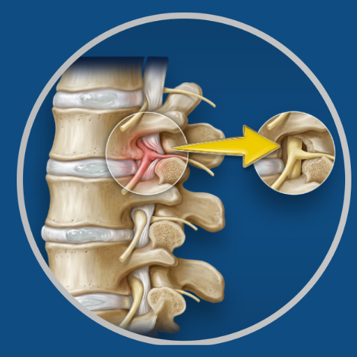
Overview
The foramen is the opening on each side of the spinal canal through which individual nerve roots pass as they exit the spine. Foraminal stenosis — narrowing of this opening by disc height loss, bone spurs, or facet joint enlargement — compresses the exiting nerve root and produces radicular pain, numbness, and weakness in the distribution of that root. Foraminotomy enlarges this opening to relieve the compression. It can be performed from the back of the spine (posterior foraminotomy) or, in cervical cases, from the front (anterior approach combined with discectomy).
Before Surgery
MRI with careful review of foraminal dimensions at the affected level. CT to assess the bony contribution to the narrowing. Correlation with clinical symptoms and neurological examination to confirm the responsible level.
During Surgery
General anaesthesia. Prone positioning for posterior approach. A small incision over the affected level. The facet joint and surrounding bone are carefully trimmed to enlarge the foramen and release the nerve root. Minimally invasive tube retractor technique is used where appropriate to minimise soft tissue disruption. Duration typically 1–2 hours.
After Surgery
Hospital stay 1–2 days. Pain managed with medication. Nerve root symptoms (arm or leg pain, numbness) typically begin to improve within days to weeks.
Recovery
Return to desk work within 3–5 weeks. Return to physical activity within 6–10 weeks. The recovery profile is similar to microdiscectomy — faster than fusion procedures.
Dr. Viswanath's Approach
Foraminotomy is performed with precise removal of bone and ligament at the foraminal entrance and exit — enough to fully decompress the nerve root without destabilising the facet joint. Navigation assistance is used where the anatomy is complex or prior surgery has altered the landmarks.
Medical Disclaimer: This information is provided for educational purposes only and does not constitute medical advice.